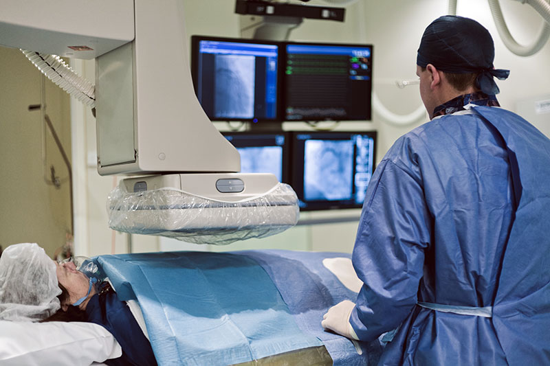
Cardiac Catheterisation, often called a coronary angiogram, is the study of coronary blood vessels and cardiac function. The heart vessels are reached through the radial (wrist) or femoral (upper thigh) artery and a contrast dye is injected to obtain images. These images help explore treatment options.
Before the procedure, a local anaesthetic is used for comfort. Since blood vessels don’t carry nerve fibres, the procedure is generally pain free. Sedation can be given if needed. Catheterisation is generally done as a day procedure.

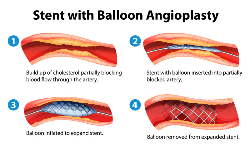
Previously termed angioplasty, the purpose of Percutaneous Coronary Interventions (PCI) is to open up blocked arteries. Similar to cardiac cath, it is performed by going through the wrist or upper thigh to insert a balloon and ultimately a stent into the blocked artery, allowing blood to flow freely. Stents are made of flexible yet strong metallic materials. Except in emergency situations, PCI is an elective procedure done separately from the initial angiogram.

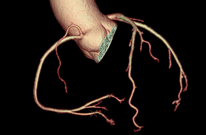
Computer tomography coronary angiography (CTCA) is a non-invasive CT scan of the coronary blood vessels although it is not as accurate as some other methods. It measures the degree of calcium deposits in the blood vessels as coronary calcium is widely recognized to be a risk factor for future heart attacks.
A CTCA is done as a quick day procedure and does not require an anaesthetic.

An Echocardiogram is the study of the heart using ultrasound, performed by placing a probe on the skin surface. Requiring no previous preparation or anaesthetic, it takes approximately 40 minutes and provides valuable information about heart function and the heart valves. An echocardiogram can be used to investigate a variety of symptoms including shortness of breath, palpitations, chest pain and loss of consciousness.

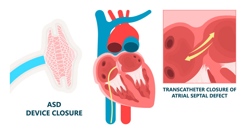
A number of structural cardiac defects can be fixed via small catheters inserted through the upper thigh. Examples include most PFO and ASD.
A PFO is a small slit like opening that connects the upper chambers of the heart. In some people, it can be associated with problems like stroke of unknown cause or even decompression illness from scuba diving. The decision whether or not to close a PFO should always be discussed with an experienced interventional Cardiologist.
ASD is a hole between the upper chambers of the heart. A large ASD can result in right heart failure leading to symptoms including breathing difficulties. The majority of ASD can now be successfully closed.
Percutaneous Structural Procedures are done under a general anaesthetic.

